Dr Sarah Bugby
Lecturer – Physics
Sarah Bugby works in the field of medical physics. Her research interests lie in detectors and imaging systems. Building on existing research and expertise, she has advanced the development of a unique handheld 3D gamma imaging device that will improve the diagnosis and treatment of a range of conditions – including cancer – and has applications beyond medicine.
Hybrid gamma camera to accelerate cancer diagnosis and treatment
Gamma cameras detect the gamma rays emitted by radiopharmaceuticals as they move through the body. The created images show function rather than anatomy – for example, highlighting changes in metabolic processes and blood flow – helping to detect and diagnose possible ill-health. Although invaluable tools within nuclear medical practice, they are expensive and a similar size to CT scanners. Reducing both their size and cost will make them more accessible to clinicians around the world.
Clinical gamma cameras are important diagnostic tools, but are large machines – taking up a whole room within a specialist nuclear medicine department. This means that even very sick or frail patients have to travel to them rather than vice versa.
A portable device would complement these invaluable machines – making them accessible to clinicians caring for patients anywhere, including intensive care units, and during radioguided surgical procedures in the operating theatre.
In 2018, I was appointed as an STFC Innovation Fellow to translate core STFC-funded research in detectors and imaging systems into a new portable device. Working with a fantastic team, I’ve been able to progress it from bench testing to clinical pilot studies – and am now supporting Serac Imaging Systems as they develop it commercially.
Developing potentially life-saving devices is a long way from where I thought I’d be when I left school. The thrill of transforming an idea into a valuable and useful application is so exciting – I love it.
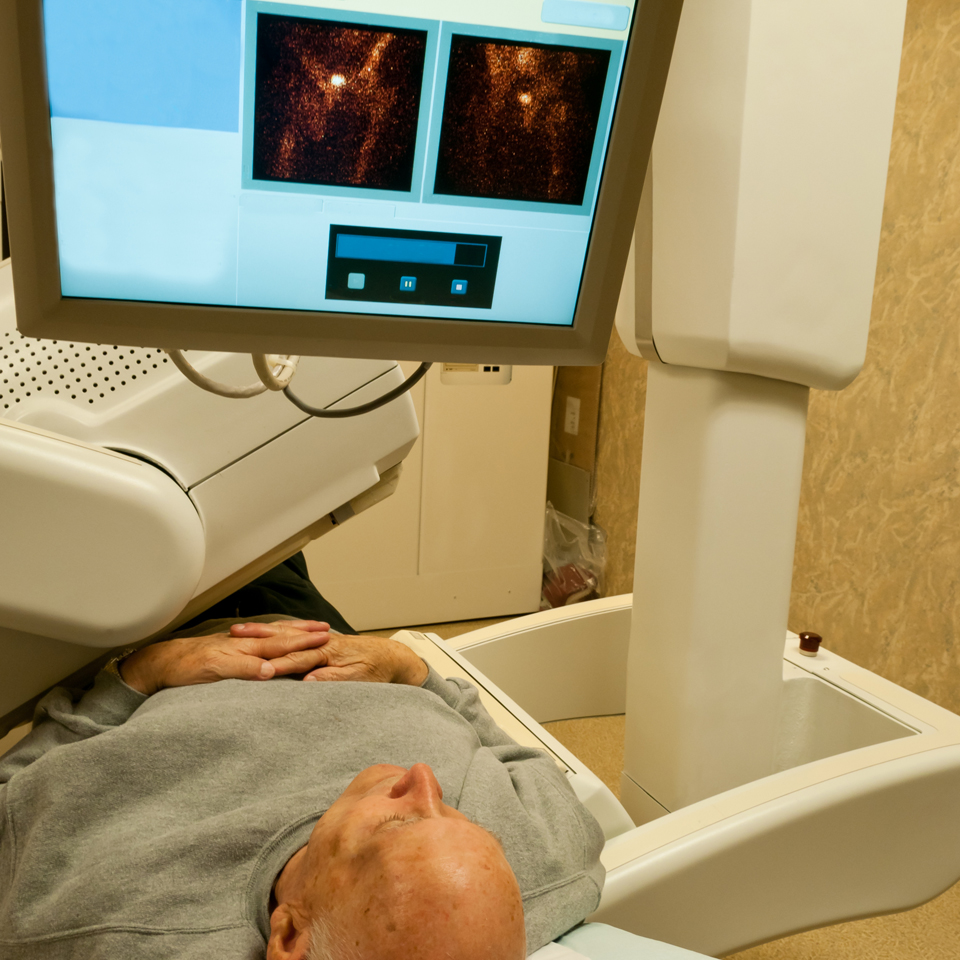
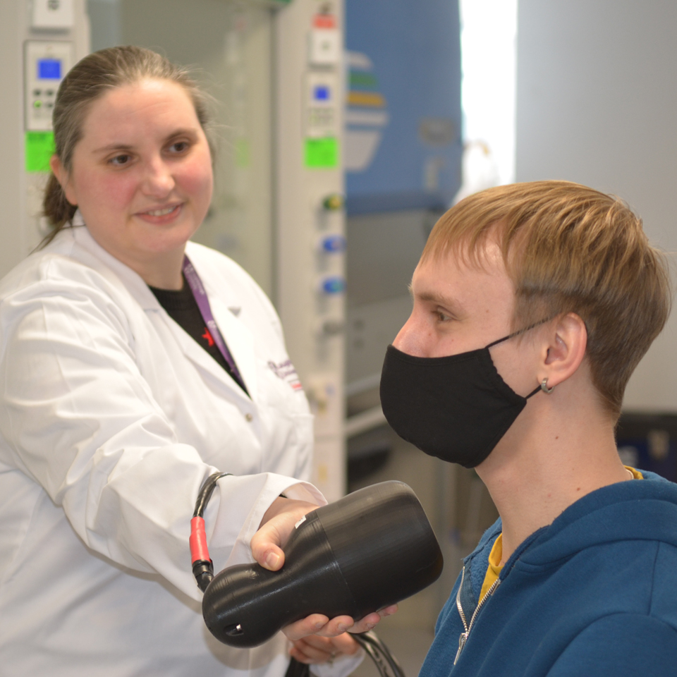
Our Hybrid Gamma Camera is about the size of a hairdryer, making it small and easy to use. The camera is positioned close to the patient’s body – for example, their neck if a thyroid problem is being investigated – and records both gamma and optical information. Clinicians can see the distribution of gamma radiation and how this relates to the patient’s anatomy which can be helpful when localising sources.
We’ve recently shown that – by taking two images from slightly different angles and triangulating the exact position of the gamma source – the camera can provide 3D information. Combining the portability of current handheld gamma imaging tools – that provide only 2D information – with the 3D capabilities of larger static machines would provide the same clinical insight in a far more flexible package.
And, of course, this can be done anywhere – not just in a specialist facility.
We’re hoping to work with colleagues in Uruguay to get the camera into the hands of physicians there. Currently, there are only three large gamma imagers in the whole country – at medical centres in Montevideo, Durazno and Salto.
At the moment, patients have to travel to one of the three centres for initial imaging, then travel home for surgery. Meanwhile, a nuclear medicine physician must travel – with a gamma probe – to support the surgeon during surgery. And if patients can’t visit one of the imaging centres, they’re likely to have more invasive surgery than necessary.
If all goes well with this partnership, we hope that we can see our portable device rolled out worldwide wherever it’s needed.
Alongside my meditech work, I’ve been part of the National Nuclear Laboratory’s Game Changers programme – developing Gamma Optical Video Imaging (GOVI) for use in Post Operational Clean Out at Sellafield.
With some modifications, our camera will help with the manual clean-up of residual radioactive material during nuclear decommissioning. Mounted on an autonomous vehicle it could also be used remotely, avoiding operative exposure to risk in unsecured or toxic areas.
Gamma imaging technology is evolving rapidly. Part of my research involves testing new types of detectors to improve sensitivity, provide more detailed information, and – using simulations and mathematical modelling – explore novel imaging techniques.
My research journey
I sometimes describe myself as the Accidental Physicist.
When I finished my A-Levels, I worked for a couple of years doing various things and realised that none of them were really for me. I decided to return to education – hoping that a university qualification might open some different employment doors for me.
I’ve always loved science, so a STEM degree seemed a logical step. The University of Leicester was easily commutable and – after a few years in my own place – I had no appetite to live in halls. So, I applied to study Physics.
During my third year, while a lot of my friends were looking forward to moving on, the process of working on research projects really clicked with me. I love looking at a problem, exploring at it from all angles, and trying to untangle it – a very necessary attribute for a researcher. I was lucky that my tutors and lecturers were fantastic mentors, and they pointed me towards postgraduate study.
So, I stayed at Leicester to embark on a PhD and then, when I graduated, secured a Postdoctoral Researcher role.
In 2019, I joined Loughborough – taking up a Lectureship and joining the Centre for Sensing and Imaging Science – continuing my work in Medical Physics. I’m happy here – the campus is beautiful, and people are welcoming and friendly.
I now co-lead the University's Applied Radiation and Medical Physics Group (ARAMP) with Jenny Spiga and Fasil Kidane Dejene. Working with researchers, clinicians and industry partners worldwide, we are developing new techniques to solve societal challenges and improve patient care.
Doing research that is helping to develop potentially life-saving devices is a long way from where I thought I’d be when I left school in 2005. The thrill of seeing an idea grow and progress into a valuable and useful application is so exciting – I love it.