CT Fracture for 3D virtual teaching
Introduction
This case study highlights the use of CT fracture imaging in the development of Mission Materials: A VR Escape Room for Extreme Temperature Material Testing, a Loughborough University 2025 DigiLabs Teaching Innovation Award (TIA). By simulating challenging testing environments in VR, the project expands access to practical materials science education—especially where real-world experimentation is limited by safety, cost, or equipment constraints. The integration of CT fracture data into VR aims to enhance student engagement, foster deeper understanding of material behaviour under stress, and develop skills in data interpretation, critical thinking, and collaborative problem-solving within a virtual lab setting.
Experimental
A 304 stainless steel sample had been fracture tested, and the resulting fracture bar was provided, and scanned using beam settings of 120kV accelerating voltage and 100µA of current.
Results
The reconstructed 3D model was successfully imported into the visualization platform, where it can be interactively manipulated. Users are able to virtually navigate through the model to examine the fracture surfaces and investigate the underlying failure mechanisms in detail.

A 3D model reconstructed using dragonfly to model a fracture face for use in VR.
Summary
CT analysis yields high-accuracy datasets that were seamlessly integrated into a virtual environment, offering a powerful tool for enhancing undergraduate education. This case study demonstrates the versatility of CT imaging in both research and teaching, highlighting its potential for application across engineering and materials science disciplines.
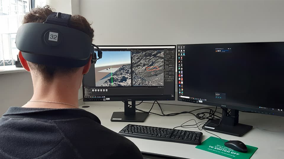 Visualising the 3D model reconstructed in the Virtual Reality environment.
Acknowledgement
Adys Yusuf, Melvin Chan & Keith Yendall – Data collection and analysis.
The DigLabs team, Dr Sina Saremi-Yarahmadi, Dr Hossein Nevisi and Dr Mark Jepson.