TEM Study of Skeletal Muscle Biopsy case study
Introduction
Skeletal muscle is a highly adaptable tissue that undergoes morphological and metabolic changes in response to exercise and training. In response to prolonged resistance training, skeletal muscle size increases, primarily due to muscle fiber hypertrophy. However, whether fiber number also changes remains controversial, and data on myofibril plasticity are limited. This study compares muscle fiber and myofibril characteristics in long-term resistance-trained (LRT) and untrained (UNT) individuals.
Experimental
Muscle biopsies were taken from the biceps brachii to measure muscle fiber area, myofibril area, and myosin spacing from volunteers. Fixed tissue was rinsed in phosphate buffer, secondary fixed in osmium tetroxide, dehydration and resin embedded prior to cross-sectioning using an ultramicrotome to prepare sections of 80 nm thickness for TEM examination. The sections were subsequently stained with uranyl acetate 2%. An FEI Tecnai F20 TEM equipped with a Gatan Orius 200 CCD camera was used to record the images.
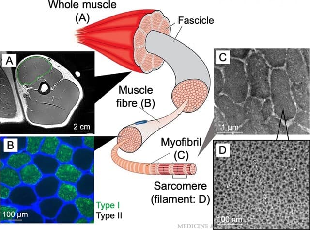
Illustration of skeletal muscle structure and obtained example images of ACSAmax of the biceps brachii (A), muscle fiber (B), myofibril (C), and myofilament (D). The arrows in D show how the myosin spacing (distance) was measured. The illustration of skeletal muscle structure was purchased from Dreamstime.com (file ID: 80735107 created by Legger) and adapted with permission.
Results
1.Muscle Growth Mechanisms – Resistance training increases skeletal muscle size, primarily through muscle fiber hypertrophy, though changes in fiber number remain controversial.
2.Differences in Trained vs. Untrained Individuals – Long-term resistance-trained individuals have larger biceps brachii muscle areas due to both greater muscle fiber size and increased fiber number. Their larger fibers contain more myofibrils, but myofibril size remains unchanged.
3.Novel Ultrastructural Adaptations – LRT individuals exhibit greater myofilament packing density, suggesting skeletal muscle ultrastructure adapts to functional overload, potentially influencing specific tension and strength.
Summary
The larger muscles of LRT individuals exhibited more fibers in cross-section and larger muscle fibers, which contained substantially more total myofibrils and more packed myofilaments than UNT participants, suggesting plasticity of muscle ultrastructure.
Medicine & Science in Sports & Exercise 56(10):p 1906-1915, October 2024. | DOI: 10.1249/MSS.0000000000003495
Acknowledgement
Dr. Zhaoxia Zhou