Polypropylene CL case study
Introduction
Cathodoluminescence (CL) allows for the high-resolution analysis of polymer blends. In this study, a polypropylene (PP) and nylon blend was analysed using scanning electron microscopy (SEM) coupled with CL to distinguish the distribution of the two polymer phases.
Experimental
Polypropylene blended with nylon was compression molded to create the sample. To prepare for SEM and CL analysis, the samples were cryo-fractured and carbon coated to prevent charging effects. CL- Panchromatic imaging was performed using a 5 keV accelerating voltage and a probe current of 1.6 nA.
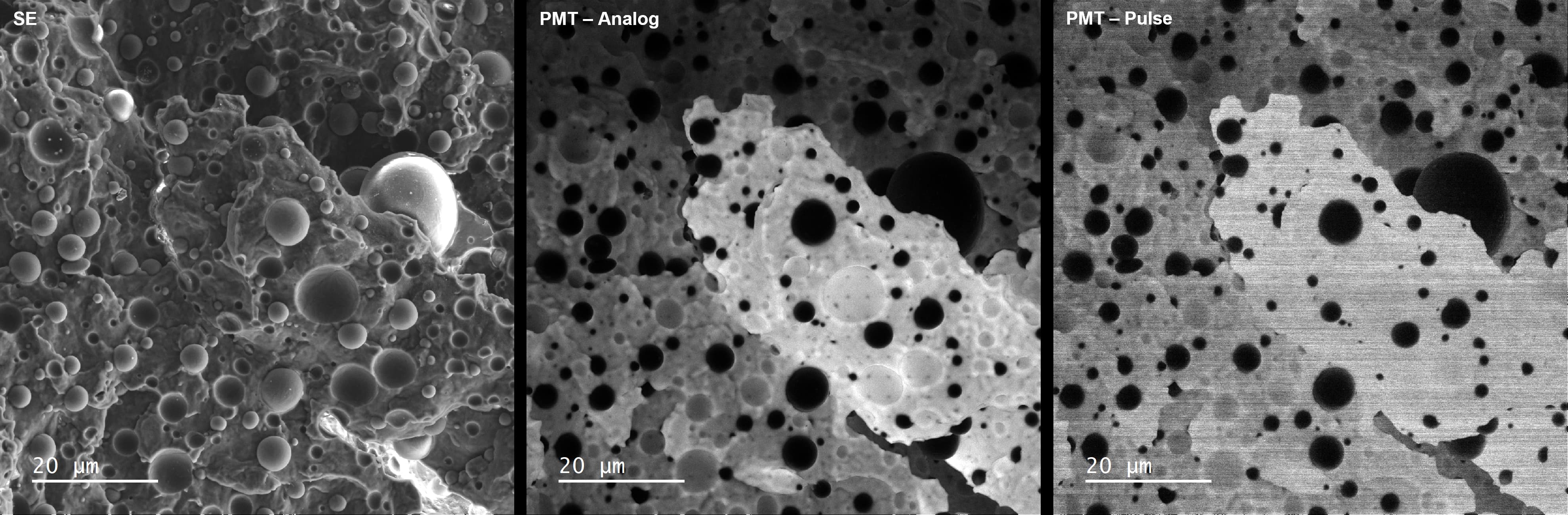 Comparison between secondary electron, PMT-Analog & PMT-Pulsed imaging modes.
Results
The microstructural analysis revealed that polypropylene formed the continuous matrix, while nylon was dispersed as spherical domains within the polymer blend. CL imaging successfully resolved these nylon spheres with minimal edge effects, enhancing their visibility compared to secondary electron imaging alone. The absence of significant edge effects in CL micrographs made this technique effective for particle size and distribution analysis.
Summary
Cathodoluminescence imaging proved to be an effective tool for analysing the phase distribution in polypropylene-nylon polymer blends. The high contrast of nylon spheres in the CL images make this approach useful for particle analysis, offering an advantage over traditional SEM techniques. This study highlights the potential of CL in polymer research, particularly for materials with distinct luminescent properties.
Acknowledgement
Stuart Robertson
Funding
The authors acknowledge access to the National Facility for High Resolution CL Analysis of Photovoltaic and Optoelectronic Devices, funded by the EPSRC (Grant No. EP/X030245/1).