Current handheld gamma imaging tools are small and easy to use, but are limited to providing 2D information, giving doctors and surgeons only part of the overall picture.
Much larger systems are able to give three-dimensional images, however, they are bulky and complex – often occupying entire rooms.
Now, researchers from Loughborough University have published a paper which shows it is possible to combine the best aspects of both devices.
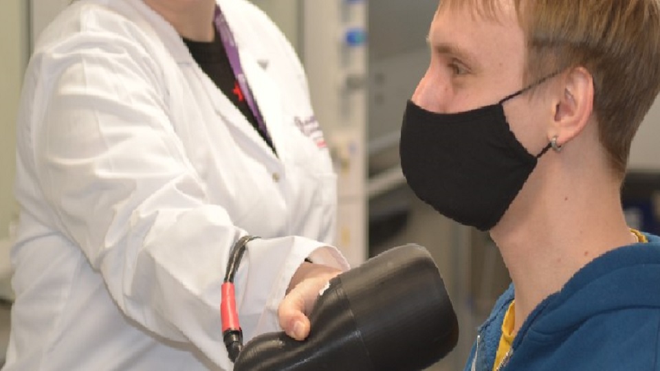
Lead author Dr Sarah Bugby, of the School of Science, is developing the Hybrid Gamma Camera (HGC) – a device about the size of a hairdryer which can easily be carried to wherever it is needed.
The original 2D HGC was created around five years ago at Leicester and Nottingham universities, where Dr Bugby joined the project before bringing the camera to Loughborough to continue the work.
It works by taking two images from slightly different angles and calculating the exact position of whatever it is observing, the exact same way astronomers measure the vast distances to stars.
Dr Bugby said: “We showed that it was possible to conduct handheld stereoscopic gamma imaging, which will provide 3D rather than 2D information.
“By combining gamma and optical imaging, this 3D information will tell the user where and how deep a source of radioactivity is inside a particular material.
“This has applications in radioguided surgery – where a surgeon is looking for a source of radioactivity within the body for example during cancer treatment and diagnosis – and may also find use in other areas in the nuclear industry.”
The University is also hoping to work with academics in Uruguay to get the camera into the hands of physicians.
The device would give medical professionals greater flexibility as there are only three large imagers in the country – at medical centres in Montevideo, Durazno and Salto – which are capable of gamma imaging.
Dr Bugby said: “Currently, a patient must travel to one of those centres, in some cases hundreds of kilometres away, for initial imaging, then travel back to their city of origin for surgery.
“A nuclear medicine physician must travel to the city where the surgery will be performed, bringing a gamma probe, in order to help the surgeon, locate the sentinel node during surgery.
“If patients cannot attend one of the nuclear medicine centers, they won’t have SLN performed and they will have all their axillary nodes removed with its associated morbidity, basically, a more invasive surgery than would otherwise be needed.”
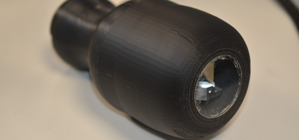
The camera works by using a pinhole (pictured above) – the small hole in the centre of the device – which allows an image of the source of gamma radiation to be taken.
Doing this twice from two slightly different positions allows the camera to triangulate the exact distance from the source giving an accurate 3D reading.
Due to the compact size of the gamma imaging technology, this could be done with a handheld system.