Research

Bioactive wound dressings based on nanofibres
The current research focuses on the development of micro- and nanotechnologies for the production of bioinspired structures with controlled porosity. Electrospinning, 3D printing and phase separation methods are used to process natural and synthetic biopolymers in order to create hierarchical and porous scaffolds for tissue engineering.
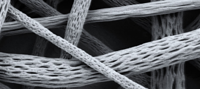
Physical and chemical traumas are often responsible for injuries of the human skin, with the consequent disruption of its normal physiology and the initiation of specific healing processes to restore the tissue functions. Acute or chronic wounds affect millions of people annually, and their incidence is expected to increase in the next years mainly due to the growth and aging of global population. Therefore, advances in wound care medical devices are required. The current research at the Materials Department of Loughborough University aims to develop wound dressings based on bioactive nanofibres. The ultrafine size of the fibres guarantees excellent conformability of the non-woven mat to the wound site and it proves complete coverage of the injured tissue and protection against infections and dehydration. Moreover, the high porosity of the meshes facilitates the transport of nutrients and the absorption of exudates, together with the effective delivery of therapeutic substances.
References
Zhang, W, Huang, C, Kusmartseva, O, Thomas, NL, Mele, E (2017) Electrospinning of polylactic acid fibres containing tea tree and manuka oil, Reactive and Functional Polymers, 117, pp.106-111, ISSN: 1381-5148. DOI: 10.1016/j.reactfunctpolym.2017.06.013.
Mascia, L, Su, R, Clarke, J, Lou, Y, Mele, E (2017) Fibres from blends of epoxidized natural rubber and polylactic acid by the electrospinning process: Compatibilization and surface texture, European Polymer Journal, 87, pp.241-254, ISSN: 0014-3057. DOI: 10.1016/j.eurpolymj.2016.12.033.
Hajiali, H, Heredia-Guerrero, JA, Liakos, I, Athanassiou, A, Mele, E (2015) Alginate Nanofibrous Mats with Adjustable Degradation Rate for Regenerative Medicine, Biomacromolecules, 16(3), pp.936-943, ISSN: 1525-7797. DOI: 10.1021/bm501834m.
Top photo: 1 µ EHT = 3.00 kV Signal A = SE2 Mag =20.00 K X WD = 11.5 mm
