
In a new study, the ‘Fourier Transform Infrared (FT-IR) microspectroscopy’ technique has been shown to identify a single cancer cell in a blood sample.
The research team – which brings together academics from University Hospitals of North Midlands NHS Trust (UHNM), Keele University, and Loughborough University – believe the breakthrough could enable doctors to monitor cancer in real time using a simple blood test.
Professor Josep Sulé-Suso, Associate Specialist in Oncology at UHNM and lead author of the study, said: “Our team was able to detect a single lung cancer cell in a patient’s blood by combining advanced infrared scanning technology with computer analysis, focusing on the unique chemical fingerprint of cancer cells.
“This approach has the potential to help patients receive earlier diagnoses, personalised treatments, and fewer invasive procedures, and it could eventually be applied to many types of cancer beyond lung cancer."
How the technique works
Circulating tumour cells (CTCs) are a type of cancer cell that can break away from a tumour and travel in the bloodstream. They can provide vital clues about how the disease is progressing and how well treatment is working. CTCs are also the cells that can lead to the spread of cancer (metastases).
Current methods for detecting CTCs can be complicated, expensive and time-consuming – and they can sometimes miss cancer cells altogether, as the cells often change their characteristics while circulating in the blood.
The research team’s method detects CTCs in a blood sample by shining an infrared beam onto it – similar to the light in a TV remote control, but far more powerful.
Different chemicals absorb infrared light in different ways, and CTCs have a distinct absorption pattern, or ‘chemical fingerprint’.
Computer analysis of the infrared absorption data can quickly identity whether circulating tumour cells are present.
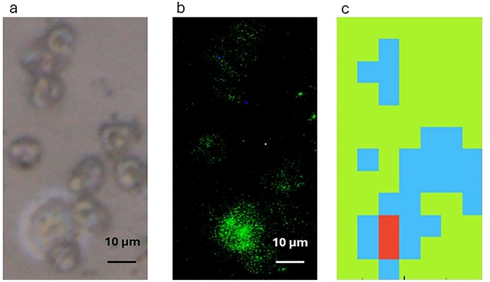
This figure shows how the technique identifies a single circulating tumour cell (CTC) in a lung cancer patient’s blood sample. The sample is first viewed under a microscope (a), and, after being tested, the cancer cell is stained (b) so the researchers can confirm it is a cancer cell. The Fourier Transform Infrared (FT-IR) microspectroscopy technique produces a colour-coded map (c) distinguishing the CTC (red) from surrounding blood cells (blue/green).
The technique is simpler and more affordable than existing approaches and uses standard glass slides already found in pathology labs to prepare blood samples for analysis under the infrared instrument, making it easier to adopt into everyday clinical practice.
Professor Paul Roach, an expert in biomaterials and interface science at Loughborough University, is part of the team that developed the method and they used the Department of Chemistry’s FTIR technology.
Professor Roach – who is also using FTIR to study microplastics in human tissues, environmental contamination, and biomaterial interactions – says this project has been especially meaningful.
“Contributing to research with the potential to transform early cancer detection is both a professional privilege and a deeply personal motivation”, said Professor Roach.
“Professionally, it offers the chance to translate fundamental spectroscopy into meaningful medical impact. Personally, it resonates strongly: cancer has affected my own family and claimed the lives of friends, and helping to expand the tools available to fight this disease gives me a powerful sense of purpose.
“The possibility that our work could one day influence clinical practice, reduce diagnostic delays, and improve patient outcomes is a constant driving force behind this research.”
What’s next?
The team will now test this method in larger patient groups, aiming to develop a rapid, automated blood test that could be integrated into NHS cancer care pathways.
They welcome collaborations with clinical, healthcare, and industry teams – to support the validation, refinement and eventual adoption of FTIR-based diagnostic tools – and research groups developing new analytical technologies, data interrogation methods, or advanced computational tools, all of which play a vital role in accelerating this work.
Read the study
The study, titled ‘Fourier Transform Infrared Microspectroscopy as a Liquid Biopsy Tool to Detect Single Circulating Tumour Cells in the Blood of a Lung Cancer Patient’ was published in the Applied Spectroscopy journal and can be read in its entirety online.
Note, the project was supported by UHNM Charity and the North Staffordshire Medical Institute, with additional backing from Keele University and Loughborough University.